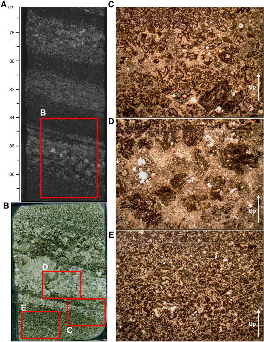
Figure F26. (A) Core section image, (B) thin section scan, and photomicrographs (plane-polarized transmitted light) of sparsely vesicular vitric tuff in Section 324-U1348A-18R-1, top of Unit III, with (C) glass shards and bubble wall fragments, (D) subrounded vesicular basalt clasts, and (E) fine hyaloclastite matrix, Hole U1348A. Thin section scan location of B is indicated by the red box in A. Width of field of view of photomicrographs is ~15 mm (1.25x) in panels C, D, and E. White arrows = orientation in core.
Previous | Close | Next | Top of page