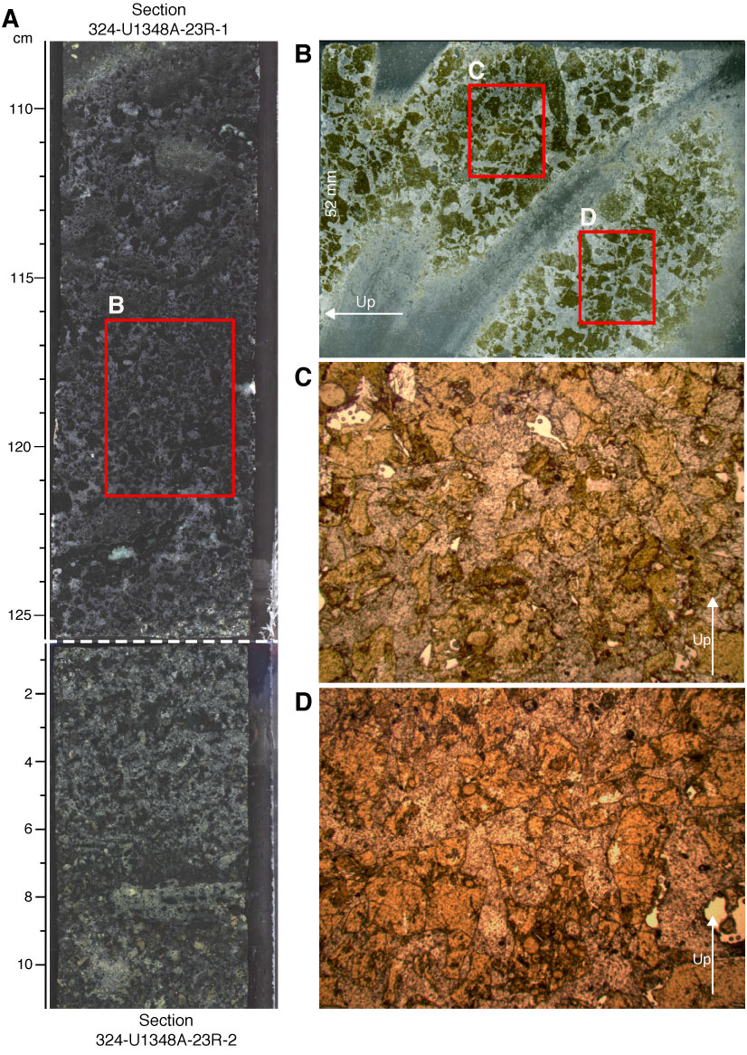
Figure F27. (A) Core section image, (B) thin section scan of angular glass shards in calcite cement, and (C, D) photomicrographs (plane-polarized light) of glass shards (bright colored fragments) at the top of Unit VI, Hole U1348A. Thin section scan location of B is indicated by the red box in A. Red boxes in B indicate photomicrograph locations. Width of field of view of photomicrographs is ~15 mm (1.25x).
Previous | Close | Next | Top of page