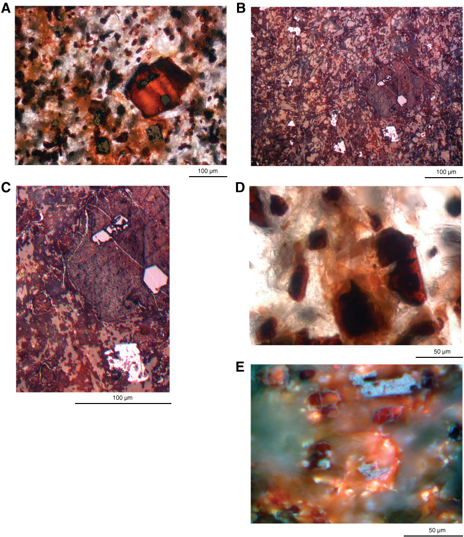
Figure F28. Photomicrographs of hematite in basalt in Unit IV, Hole U1349A (Thin Section 222; Sample 324-U1349A-10R-3, 97–99 cm). A, B. Altered olivine enclosing two Cr spinel crystals, light gray in B. The numerous specks in the majority of the photomicrograph are variably crystalline iron oxyhydroxides and goethite(?)-hematite. In B, hematite lines small cracks crossing the altered olivine and outlines rims of some of the groundmass oxyhydroxides. C. Same as B at higher magnification. D. Hematite partly replacing altered clinopyroxene. E. High-magnification view of groundmass oxyhydroxides with hematite rims. Much of the view did not polish and so is outside of the restricted focal range of the objective and is therefore blurry. Two skeletal titanomagnetite crystals are gray with fine exsolution lamellae; the lower one is centered on a crystal that is almost completely transformed to hematite-oxyhydroxide (red internal reflections surrounding the gray). A is under transmitted with partially reflected light; B and C are under reflected light; D is under transmitted light; E is under oil-immersion reflected light.
Previous | Close | Next | Top of page