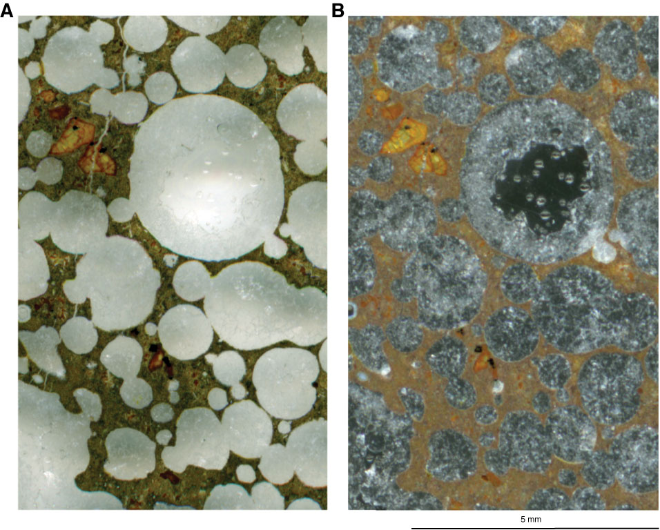
Figure F29. Thin section images using different techniques (Thin Section 211; Sample 324-U1349A-7R-4, 32–35 cm). A. White background showing rock contrasted with filled vesicles. Altered olivine with spinel occurs at the upper left and lower center middle. B. Scanning light passed up through the section to a dark background. This reveals outlines of the infilling calcite, subduing the image of the rock itself except to show how strongly stained it is with iron oxyhydroxides.
Previous | Close | Next | Top of page