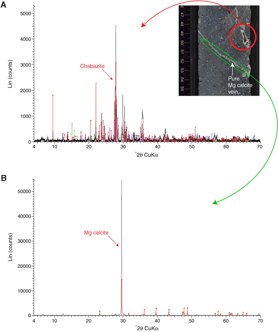
Figure F23. X-ray diffraction spectra and associated core photograph (Sample 330-U1373A-8R-4, 48–52 cm). A. Vugs and vesicles showing filling with zeolites (chabazite and phillipsite) and nontronite (blue coating of vesicles). Blue lines = phillipsite, green lines = nontronite. Red circle = analyzed zone. B. Vein predominantly composed of Mg calcite. Green oval = analyzed zone.
Previous | Close | Next | Top of page