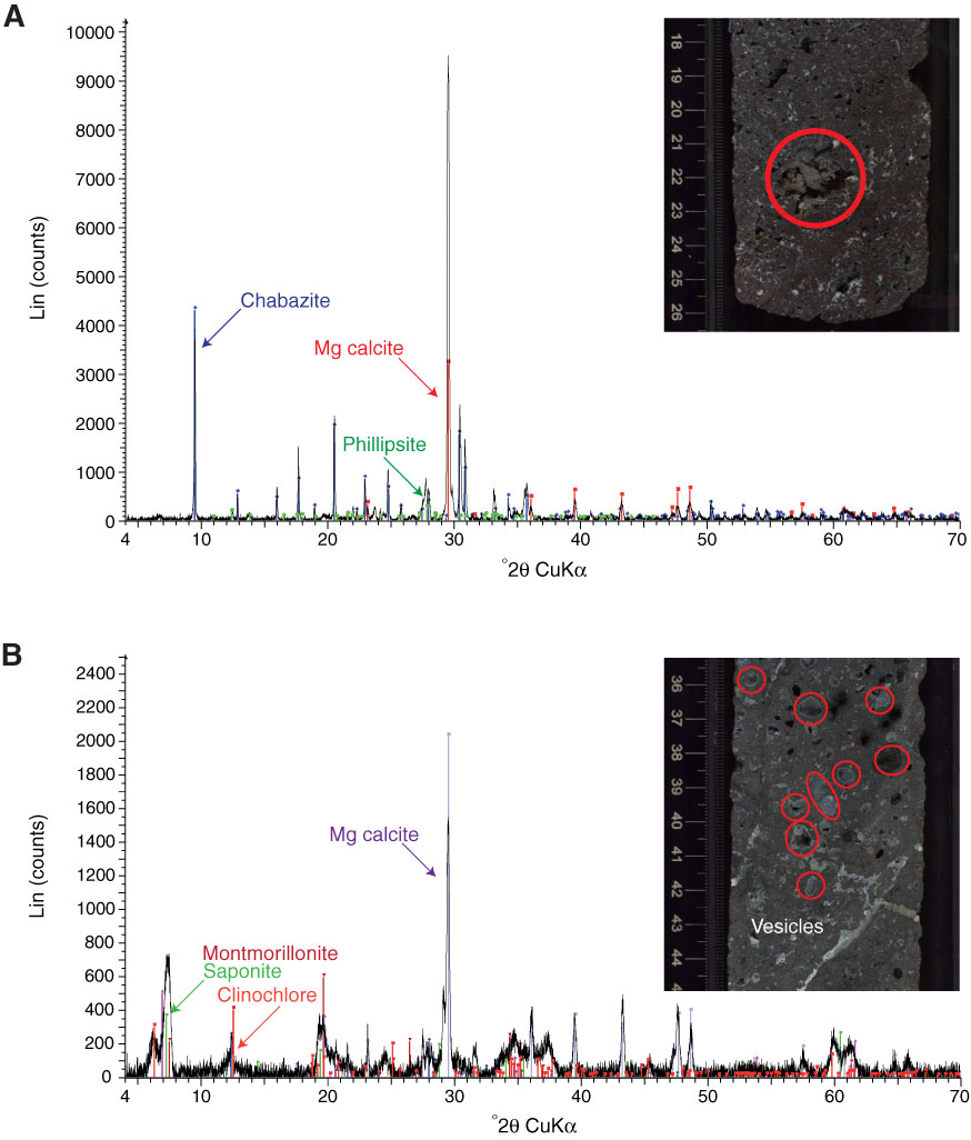
Figure F26. X-ray diffraction spectra and associated core photographs (red circles = analyzed zones). A. Vug (Sample 330-U1373A-8R-2, 22–24 cm) filled with Mg calcite and zeolite (chabazite and phillipsite). B. Vugs (Sample 330-U1373A-7R-4, 36–41 cm) filled with Mg calcite, zeolite (phillipsite, not labeled), clay minerals (montmorillonite and saponite), and small proportions of clinochlore.
Previous | Close | Next | Top of page