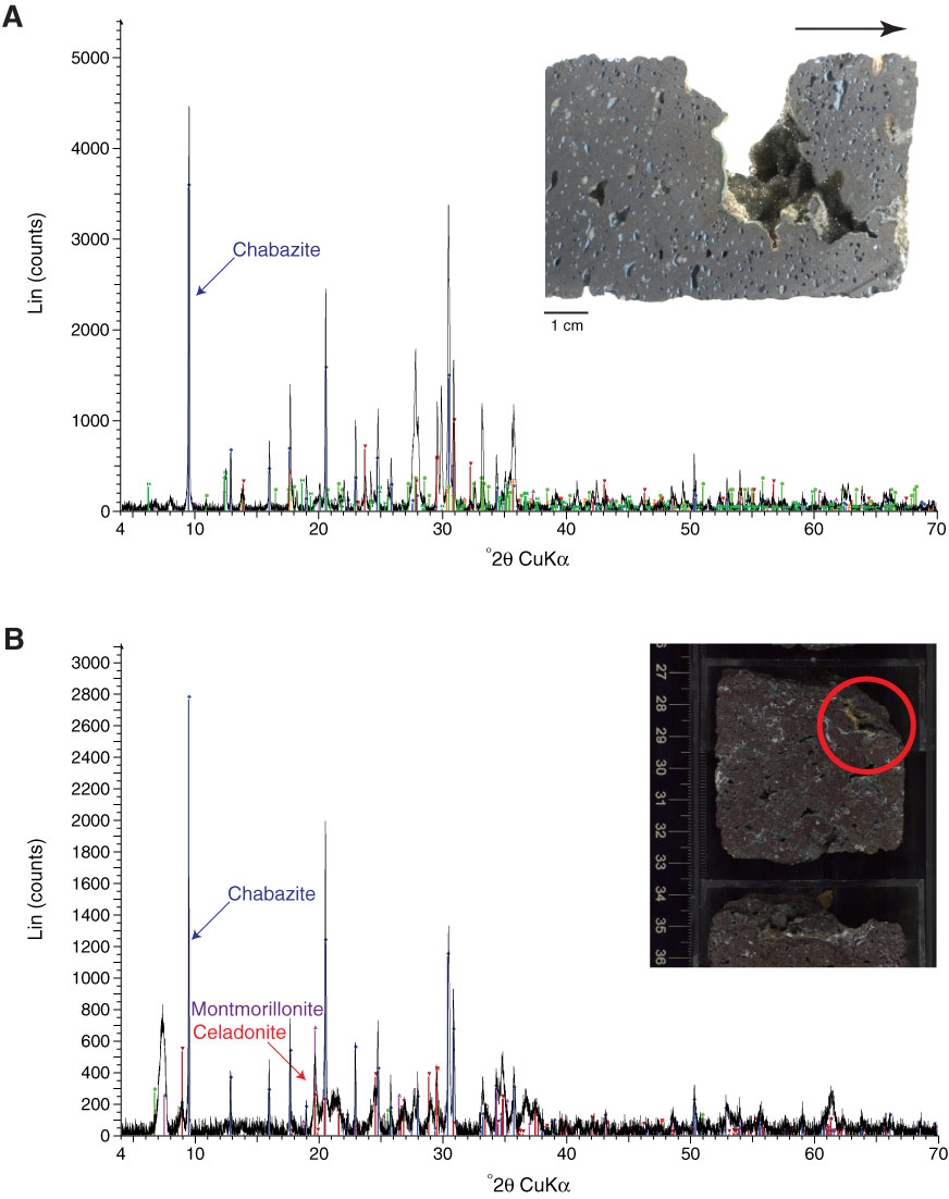
Figure F25. X-ray diffraction spectra and associated close-up photographs. A. Vug (Sample 330-U1373A-9R-1, 102–103 cm) partially filled with Mg calcite, zeolite (chabazite and phillipsite), and clay minerals (nontronite) (Mg calcite, phillipsite, and nontronite not labeled). Arrow points to core top. B. Vug (Sample 330-U1373A-8R-2, 29–31 cm) partially filled with Mg calcite, zeolite (chabazite), and clay minerals (nontronite, montmorillonite, and celadonite) (Mg calcite and nontronite not labeled). Red circle = analyzed zone.

Previous | Close | Next | Top of page