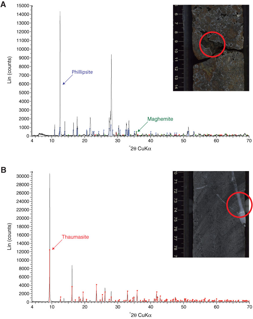
Figure F35. X-ray diffraction spectra and associated core photographs (analyzed zones are highlighted with red circle). A. Vug showing filling with zeolite (phillipsite), Mg calcite (not labeled), and maghemite (Sample 330-U1374A-25R-2, 8–10 cm). B. Vein showing filling with thaumasite (Sample 330-U1374A-59R-6, 74–76 cm).
Previous | Close | Next | Top of page