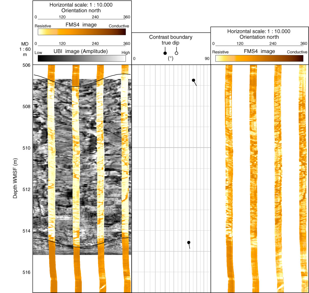
Figure F94. Summary figure of Formation MicroScanner (FMS) images showing lithologic Unit 146. Ultrasonic Borehole Imager (UBI) image is shown in gray scale, overlain by static-normalized FMS image. Top and bottom contact dip angle and direction are shown by black sinusoids and their associated tadpoles (note that direction of tadpole tail indicates true azimuth of dip, and angle of dip is where circle is plotted). FMS dynamic-normalized image is shown on right.

Previous | Close | Next | Top of page