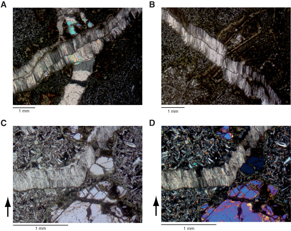
Figure F35. Representative thin section photomicrographs of veins, Hole U1376A. A, B. Early carbonate vein containing uniform crystals, crosscut by a later vein containing cross fibers (Sample 330-U1376A-6R-4W, 119–122 cm; Thin Section 252). This thin section is not oriented, so it is not possible to determine the direction of vein opening. In B the vein containing cross fibers crosscuts an altered olivine crystal. The two sides of the olivine crystal have been displaced in the same orientation as mineral fiber growth. C, D. Oriented thin section (black arrow points toward top of core) from upper part of the 33.11 m thick olivine and pyroxene-phyric flow in Unit III (Sample 330-U1376A-8R-6W, 136–140 cm; Thin Section 255): (C) plane-polarized light, (D) crossed polars. The direction of mineral fiber growth shows the direction of vein opening (Ramsay and Huber, 1987) and indicates the vein opened in two pulses, one with a near-vertical displacement and a later opening in a slightly inclined direction.
Previous | Close | Next | Top of page