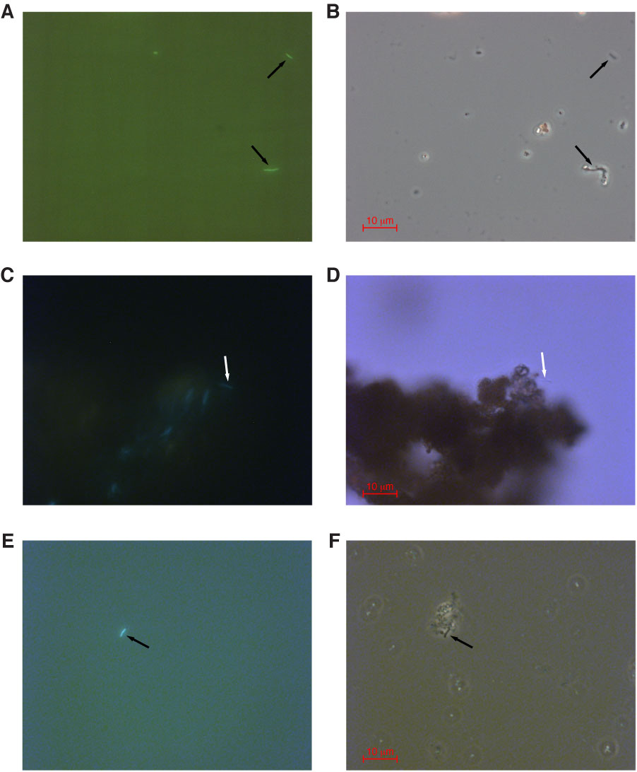
Figure F16. Paired photomicrograph images using epifluorescent (A, C, E), phase contrast (B, F), and brightfield (D) microscopy. Arrows indicate the location of putative FeOB. A, B. Section 331-C0015B-1H-1, ASW media A. C, D. Section 331-C0015C-1H-3, ASW media A. E, F. Section 331-C0015C-1H-3, ASW media B.
Previous | Close | Next | Top of page