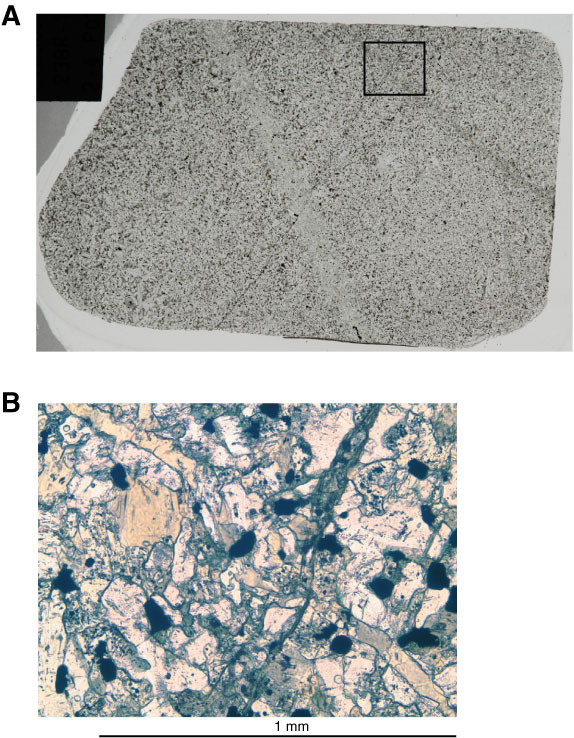
Figure F50. Veins (Sample 335-1256D-238R-1, 2–4 cm [Piece 1]; Thin Section 6). A. Photograph of granoblastic recrystallized vein (gray, left) and amphibole vein (dark, top middle to middle right) that are both cut by micrometer-sized amphibole veins. B. Photomicrograph of crosscutting relationship between a hornblende vein and a micrometer-sized branching actinolite vein (close-up of A; plane-polarized light).
Previous | Close | Next | Top of page