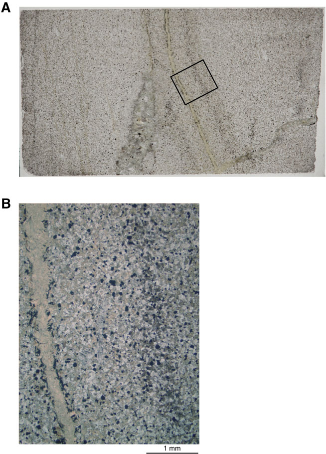
Figure F51. Photographs of amphibole veins (Sample 335-1256D-Run12-RCJB-Rock B; Thin Section 21). A. Amphibole vein with a layered alteration halo, adjacent to a felsic intrusion (bottom center) that tapers upward in photo to an amphibole vein. B. Amphibole vein lined with acicular magnetite crystals (close-up of A; plane-polarized light). In the alteration halo along the vein, pyroxenes are replaced by amphibole close to the vein, and farther from the vein clinopyroxenes are altered to dusty clinopyroxene.

Previous | Close | Next | Top of page