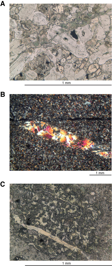
Figure F52. Photomicrographs of veins. A. Micrometer-sized amphibole vein (Sample 335-1256D-235R-1, 11–12 cm [Piece 1]; Thin Section 2) (plane-polarized light). The vein both cuts and follows grain boundaries. Where clinopyroxene crystals are bisected, most of the crystal is replaced by amphibole. B, C. Sample 335-1256D-236R-1, 0–4 cm (Thin Section 4): (B) prehnite vein with minor actinolite needles (cross-polarized light), (C) poikiloblastic epidote (1 mm) enclosing small pyroxene crystals (plane-polarized light).
Previous | Close | Next | Top of page