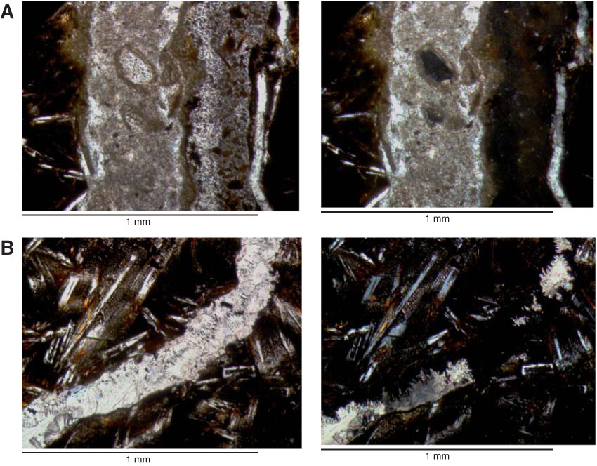
Figure F17. Photomicrographs of composite zeolite and carbonate veins. Left images are under single-polarized light; right images are under cross-polarized light. A. Photomicrograph of 1 mm wide vein with three domains (Sample 336-U1383C-2R-2, 15–18 cm [Piece 3], Thin Section 28). From right to left: (1) recrystallized calcite rim (50 µm across), (2) zeolite filling with very low birefringence and mixed with altered glass shards and Fe oxyhydroxides, and (3) micritic filling with altered glass and zeolite clasts. Field of view (FOV) = 1.2 mm. B. Photomicrograph of 0.1 mm composite vein with drusy acicular to fibrous zeolite druses with very low birefringence overgrowth by fibrous calcite (Sample 336-U1383C-29R-1, 58–64 cm [Piece 14], Thin Section 55). Later stage granular calcite showing radial extinction fills the remaining open space. FOV = 1.1 mm.
Previous | Close | Next | Top of page