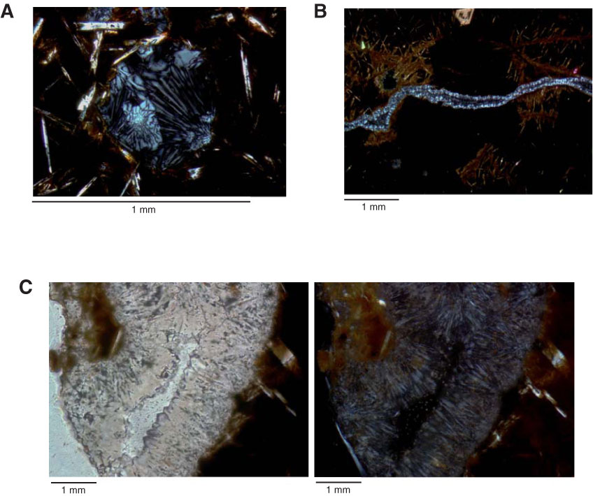
Figure F16. Photomicrographs of zeolite occurrence in veins and vesicles. A. Zeolite-filled vesicle in pervasively altered microcrystalline basalt showing radial acicular aggregates (Sample 336-U1383C-7R-2, 95–96 cm [Piece 16], Thin Section 38). Field of view (FOV) = 1.2 mm; cross-polarized light. B. Thin (0.1 mm) zeolite veinlets in variolitic cryptocrystalline basalt with interconnected hairline veinlets lined by incipient alteration halos (Sample 336-U1383C-16R-2, 30–33 cm [Piece 6], Thin Section 47). Euhedral olivine (top) remains unaltered thorough the mesostasis. FOV = 5 mm; cross-polarized light. C. Vein filled by acicular zeolite with low birefringence (Sample 336-U1383C-6R-1, 100–104 cm [Piece 17], Thin Section 34). Left image under single-polarized light; right image under cross-polarized light. FOV = 5 mm.
Previous | Close | Next | Top of page