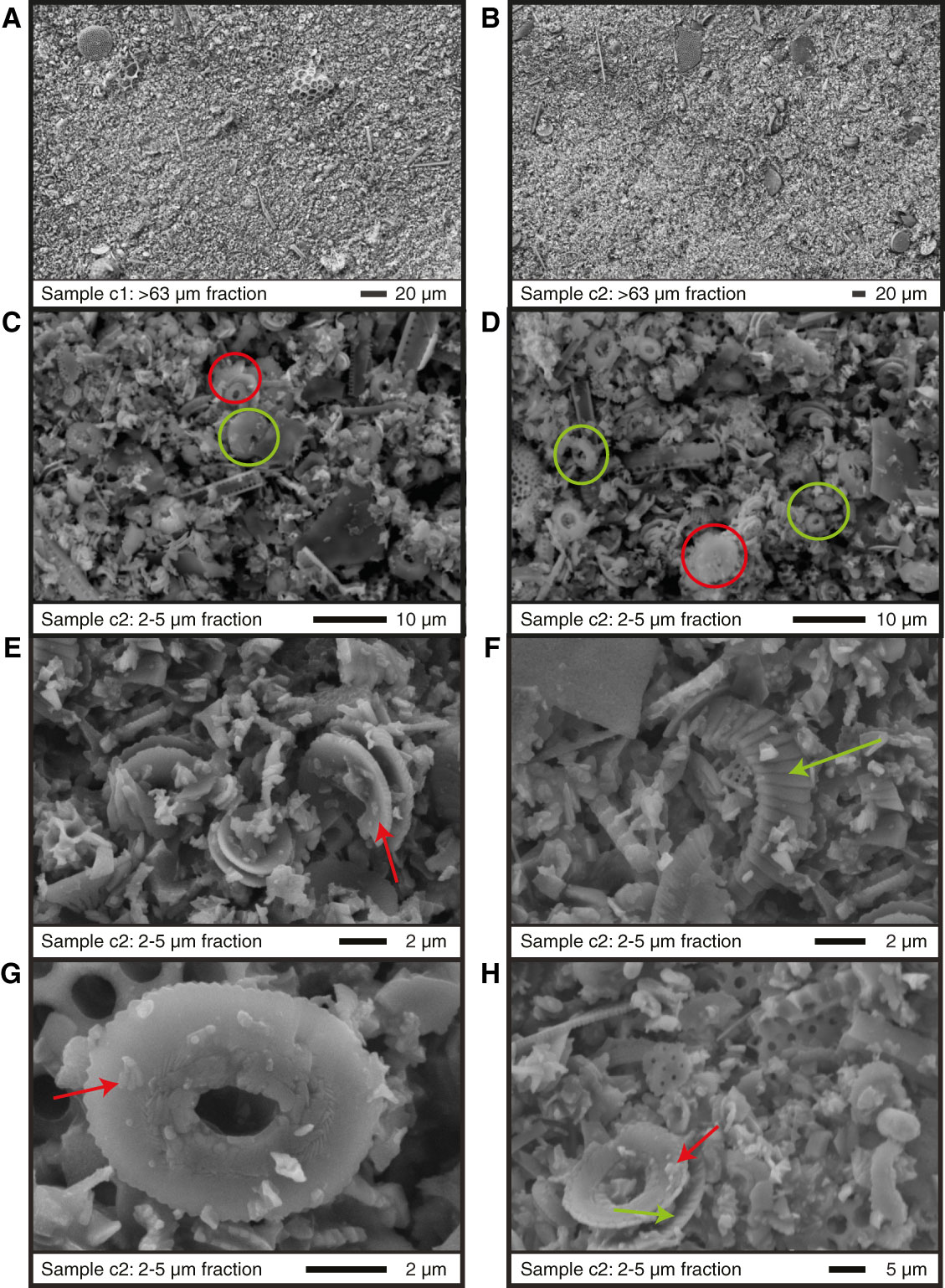
Figure F5. The BSE SEM images of the fine fraction (<63 µm) Samples (A) c1 and (B) c2 show the presence of many heterococcolith plates, whole diatoms, and large fragments of radiolarians and foraminifers. C, D. High-resolution SE SEM images of Sample c2 show the presence of large numbers of coccoliths (green circles). The images also show the presence of small fragments that are partially made up of foraminiferal and coccolith fragments (red circles). E, F, G, H. High-resolution SE SEM images of Sample c2 show that coccoliths have experienced slight to minor etching, although there is considerable fragmentation. Some original microstructures are retained (green arrows), but there is some evidence of calcite overgrowths (red arrows).

Previous | Close | Next | Top of page