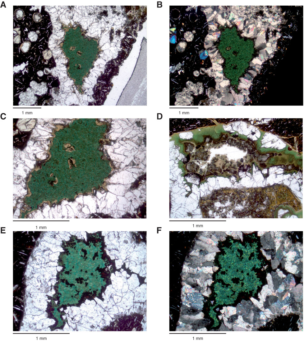
Figure F45. Thin section photomicrographs of vesicles initially filled with carbonate followed by brown (saponite, montmorillonite?) and then green (nontronite, celadonite) minerals. A–C. Sample 330-U1374A-44R-2, 121–125 cm (Thin Section 196): (A, C) plane-polarized light, (B) crossed polars. D. Sample 330-U1374A-45R-7, 22–24 cm (Thin Section 198; plane-polarized light). E, F. Sample 330-U1374A-47R-1, 133–135 cm (Thin Section 200): (E) plane-polarized light, (F) crossed polars.
Previous | Close | Next | Top of page