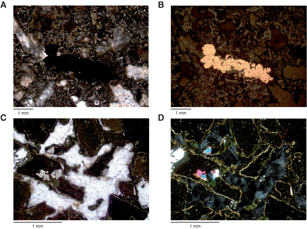
Figure F48. Thin section photomicrographs of veins and voids. A, B. Millimeter-thick vein (aphyric basalt, intrusive sheet) filled with calcite and pyrite (Sample 330-U1374A-58R-4, 28–32 cm; Thin Section 214): (A) transmitted light (plane-polarized), (B) reflected light. C, D. Voids (moderately plagioclase-augite-olivine-phyric basalt breccia) filled with zeolite (Sample 330-U1374A-49R-1, 92–94 cm; Thin Section 206): (C) plane-polarized light, (D) crossed polars.

Previous | Close | Next | Top of page